BIOMIMESYS® Liver Webinar
Applications for 3D in vitro models with BIOMIMESYS® Liver 3D cell culture systems have recently emerged as promising tools for reproducing the cellular environment and the organization of tissues/organs, where cells are connected to each other and to the surrounding extracellular matrix (ECM). In this webinar we will focus on BIOMIMESYS® Liver and will discuss […]
BIOMIMESYS® 3D cell culture scaffold as powerful tool to study metastases of breast cancer
Exciting news! A groundbreaking study using BIOMIMESYS® for breast cancer metastasis research has been published in the Experimental Hematology & Oncology Journal. The study, titled “ProNGF promotes brain metastasis through TrkA/EphA2 induced Src activation in triple negative breast cancer cells” by Cicero, Trouvilliez, et al. (2023), emphasizes the role of Extracellular Matrix organ specificity in […]
BIOMIMESYS® Adipose Tissue Webinar
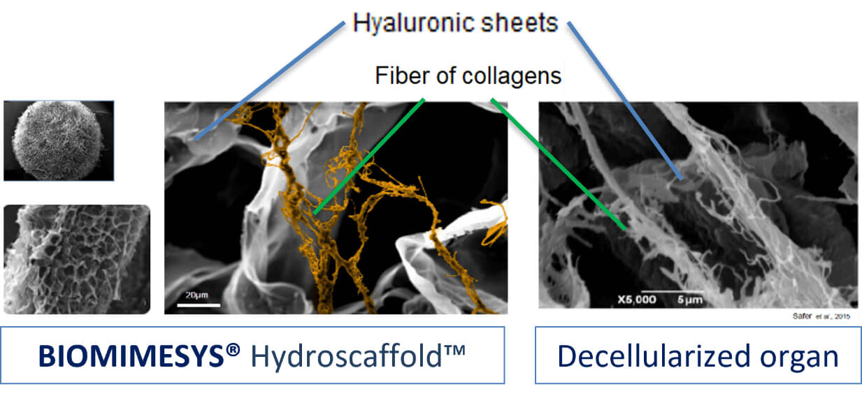
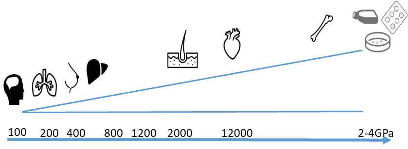
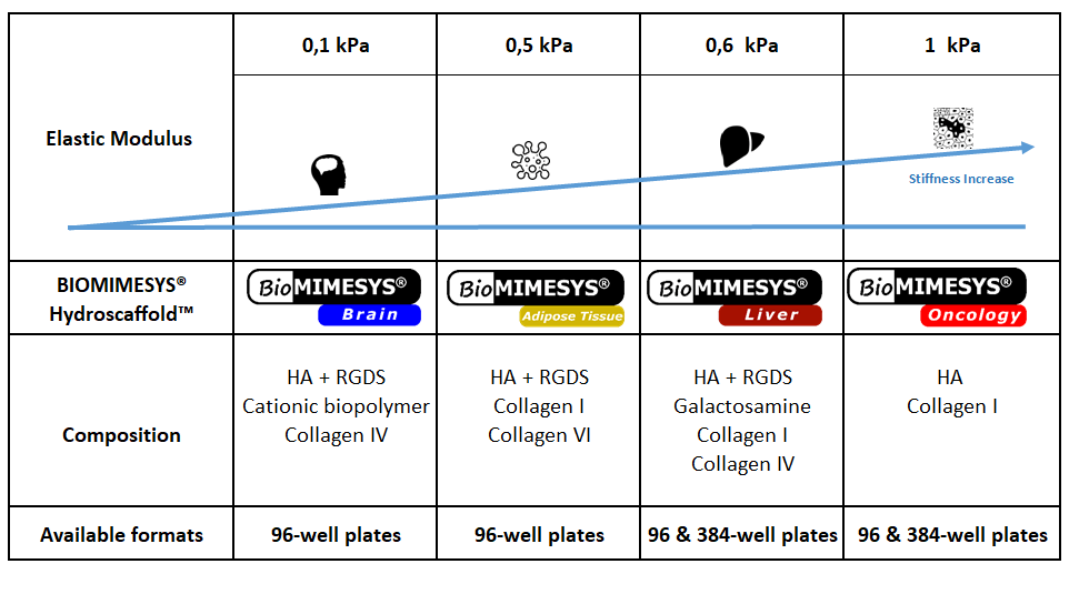
BIOMIMESYS® hydroscaffold technology based on crosslinking Hyaluronic Acid (HA) and extracellular matrix compounds reproduces tissue microenvironment in 3D. In this webinar we will focus on BIOMIMESYS® Adipose Tissue and its applications. Due to the tunable mechanical and chemical properties, this matrix possess biophysical characteristics similar to natural tissue and represent highly effective matrice for the […]
BIOMIMESYS® Webinar
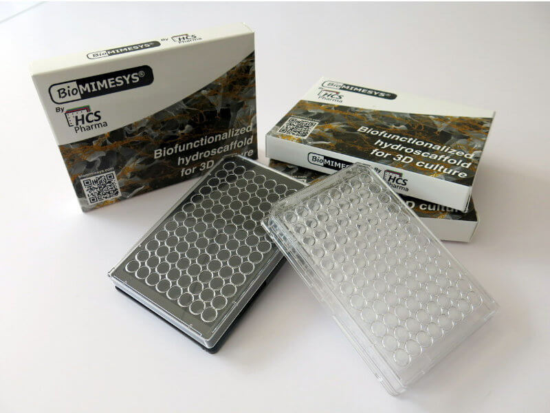
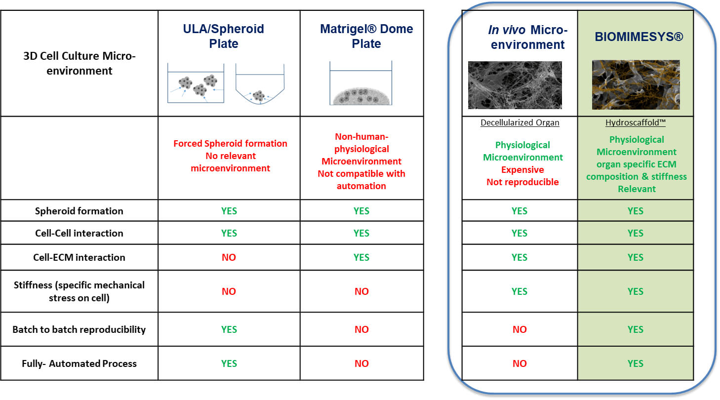
How to create physiologically-relevant 3D in vitro models using BIOMIMESYS® hydroscaffold BIOMIMESYS® is a Hyaluronic acid (HA) based scaffold 3D cell culture model. This groundbreaking 3D cell culture technology associates the behavior of a solid scaffold and of a hydrogel, which we “Hydroscaffold”. This Hydroscaffold along with its patented technology can biomimic different cell’s extra-cellular […]