It is a long established fact that a reader of a page when looking at its layout.

    BIOMIMESYS® Adipose Tissue

    3D cell culture model for adipose tissue

    Product Basics

    BIOMIMESYS® Adipose Tissue is a Hyaluronic acid (HA) based scaffold 3D cell culture model. This groundbreaking 3D cell culture technology associates the behavior of a solid scaffold and of a hydrogel, which we call “Hydroscaffold”. The Hydroscaffold’s composition and mechanical properties can mimics the microenvironment of adipose tissue. It is proven to provide better in vitro/in vivo correlation and cell longevity than 2D models.

    Key Features

    Technical Information

    Mimics the microenvironment of adipose tissue

    BIOMIMESYS® Adipose Tissue composition and organization are similar to in vivo adipose tissue by including collagens I, VI and fibronectin (RGDS pattern) in a cross-linked hyaluronic acid gel.

    BIOMIMESYS® Adipose tissue and decellularized human adipose tissue in vivo
    BIOMIMESYS® Adipose tissue (left) and decellularized human adipose tissue in vivo (5) (right)
    SEM picture of BIOMIMESYS® Adipose tissue
    SEM picture of BIOMIMESYS® Adipose tissue (collagen chains are artificially stained in gold)

    In order to demonstrate the induction of adipogenesis by the HCS Pharma hydroscaffold, BIOMIMESYS® Adipose tissue, various tests were performed on two cell types: 3T3-L1 and Human White Preadipocyte (HWP) according to the following culture protocol.

    Protocol: as in 2D culture, seeding and differentiation steps are induced from day 0 (for HWP and 3T3-L1) until complete maturation.

    Figure: A and B: Adipocyte schematic morphology in vivo.

    C and D: Light-microscopy images of adipocyte differentiated from preadipocyte HWP (Human white preadipocytes) at 7 days after seeded. HWP cultured in 2D exhibited a fibroblastic appearance (C), whereas in BIOMIMESYS® Adipocyte, it was resembling morphology of mature adipocytes in vivo, showing the establishment of their principal function, fat accumulation, resulting in the presence of vesicles (D).

    E and F : SEM images of 3T3-L1 (murine preadipocytes) at day 7 (E) and HWP at day 14 (F), both were cultured in BIOMIMESYS® Adipocyte. In the hydroscaffold, adipocytes formed aggregates which increased in size over time.

    Better differentiation and cell longevity

    BIOMIMESYS® Adipose Tissue is dedicated to the culture of adipocytes: compared to 2D culture, primary human pre-adipocytes in BIOMIMESYS® Adipose Tissue (3D) showed optimal differentiation into adipocytes, and their functionality (such as lipid storage in a single large vacuole) was maintained until 28 days.

    For a better in vitro/in vivo correlation

    Figure 2: Lipid accumulation in 3T3-L1 with or with Retinoic acid, compared to 2D cultures (n=3, two-ways ANOVA statistics with Tukey post-hoc ; * : p<0,05 ; ** : p<0,01 ; *** : p<0,001)

    To quantify the inhibition of lipogenesis, human pre-adipocytes and 3T3-L1 cells cultured in BIOMIMESYS® Adipose tissue and in 2D culture are treated with Retinoic acid, a well-known regulator. Lipid accumulation is measured using the AdipoRed ™ kit, normalized by DNA level.

    As low as 1μM of Retinoic acid led to a significant lipids accumulation inhibition in 2D, whereas at least 5μM of this molecule was needed to significantly inhibit the lipogenesis of adipocytes grown in BIOMIMESYS® Adipose tissue.

    Ready to Use

    BIOMIMESYS® Adipose Tissue is EASY & READY TO USE. Upon receiving the vacuum sealed 96-well plate open it (under a hood) and add the cells directly on top of the matrix. Changing the culture medium is easy as well. To remove medium, simply draw the medium with a pipette between the matrix and the edge of the well. To refresh the medium, place fresh medium onto the surface of the matrix.

    Successfully Tested Cells

    Cell Lines
    Human breast adenocarcinoma : MCF-7
    Human breast carcinoma : CAL-51 / T-47D / MDAMB-231 / MCF10A
    Normal mouse preadipocytes : 3T3-L1 / 3T3-F442A
    Human colorectal adenocarcinoma : HCT 116
    Normal human Keratinocytes : HaCaT
    Normal human Fibroblasts : BJ

    Primary Cells
    Human white pre-adipocyte subcutaneous : HWP cryopreserved / HPrAD
    Human Fibroblasts : NHDF
    Human Keratinocytes : NHEK

    Stem Cells
    Human induced pluripotent stem cells : hiPS
    Human sebocytes from induced pluripotent stem cells : PCi-SEB
    Complete List of Tested Cells [PDF]

    * Proof of Concept:
    In Vivo-like Morphology of Adipocytes [PDF]
    Increased Expression of Differentiation Genes [PDF]
    Higher Lipogenesis Activity [PDF]
    Maturation of Functional Human Adipocytes [PDF]
    Application: Regulation of Lipid Accumulation [PDF]

    Compatible with Most Downstream Applications

    BIOMIMESYS® hydroscaffold has many properties (transparent, porous, biodegradable, & solid) making it ideal for use with numerous downstream applications as shown below.

    Specification

    Composition:

    • Hyaluronic acid (HA) (1.6MDa) grafted with RGDS
    • Adipic acid dihydrazide (ADH) as crosslinker
    • Collagen Type I & VI

    Physicochemical Features:

    • Young’s modulus: ~0.5kPa – different stiffness is available through customization
    • Porosity: 120±50µm

    Pricing

    BIOMIMESYS®

    BIOMIMESYS® Adipose Tissue

    24 Hydrogel / 96 Well

    Transparent Plate
    • SKU: BIO_ADI_96_24_transp
    • Price: $410.00 → $369.00
    Black Plate
    • SKU: BIO_ADI_96_24_black
    • Price: $410.00 → $369.00

    96 Hydrogel / 96 Well

    Transparent Plate
    • SKU: BIO_ADI_96_96_transp
    • Price: $590.00 → $531.00
    Black Plate
    • SKU: BIO_ADI_96_96_black
    • Price: $590.00 → $531.00

    References & Literature

    1. Louis, F. et al. A biomimetic hydrogel functionalized with adipose ECM components as a microenvironment for the 3D culture of human and murine adipocytes. Biotechnol Bioeng 114, 1813–1824 (2017) doi: 10.1002/bit.26306.

    2. Neels, J. G., Thinnes, T. & Loskutoff, D. J. Angiogenesis in an in vivo model of adipose tissue development. FASEB J 18, 983–985 (2004) doi: 10.1096/fj.03-1101fje.


    3. Cristancho, A. G. & Lazar, M. A. Forming functional fat: a growing understanding of adipocyte differentiation. Nat Rev Mol Cell Biol 12, 722–734 (2011) doi: 10.1038/nrm3198.

    4. Dalby, M. J., Gadegaard, N. & Oreffo, R. O. C. Harnessing nanotopography and integrin-matrix interactions to influence stem cell fate. Nat Mater 13, 558–569 (2014) doi: 10.1038/nmat3980.


    5. McBeath, R., Pirone, D. M., Nelson, C. M., Bhadriraju, K. & Chen, C. S. Cell shape, cytoskeletal tension, and RhoA regulate stem cell lineage commitment. Dev Cell 6, 483–495 (2004) doi: 10.1016/S1534-5807(04)00075-9.


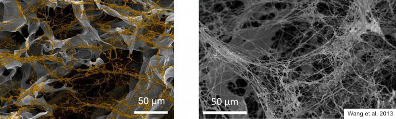
    6. Wang, L., Johnson, J. A., Zhang, Q. & Beahm, E. K. Combining decellularized human adipose tissue extracellular matrix and adipose-derived stem cells for adipose tissue engineering. Acta Biomater 9, 8921–8931 (2013) doi: 10.1016/j.actbio.2013.06.035.

    FOR RESEARCH USE ONLY, NOT FOR USE IN DIAGNOSTIC PROCEDURES