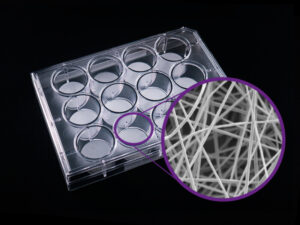
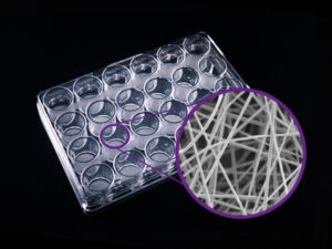
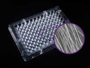
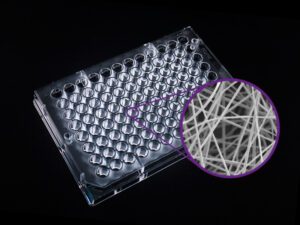
Cellevate 3D NanoMatrice™
Nanofiber scaffold for 3D cell culture
Product Basics
Nanofibers, usually defined as fibers with diameters on the nanoscale (1-1000 nm), have garnered considerable attention in both academia and the industry over the last decade. Their large surface-to-volume ratio endows them with several interesting properties that are desirable for a variety of applications. Although this type of material and its applications are still in their infancy, both the industry and academia have begun to explore its potential, making significant advancements within well-established industries.
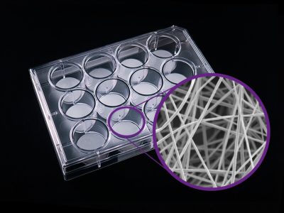
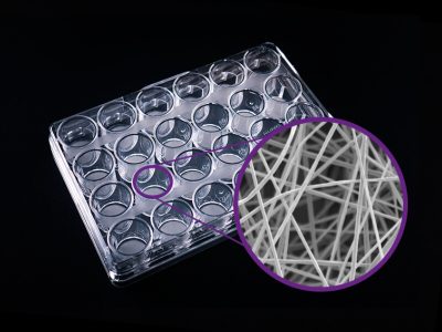
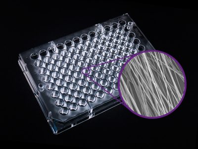
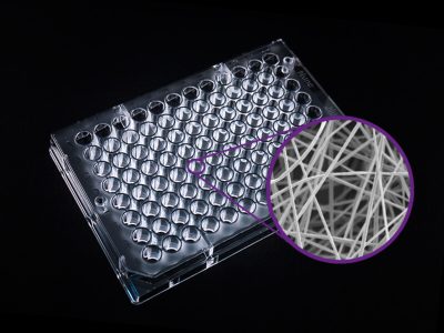
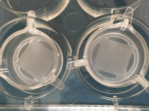
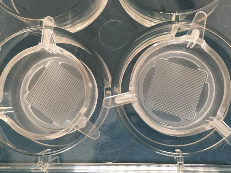
The Cellevat3d™ NanoMatrice™ is a nanofiber scaffold created from fully synthetic polycaprolactone (PCL). It is produced through a patented technique that refines the standard electrospinning method commonly used, enhancing scaffold parameter control — and consequently, the overall fiber quality — and increasing batch-to-batch consistency. It has a high specific surface area (>5m²/g) resulting in an exceptional surface-to-volume ratio, in turn increases the total biomass that can be maintained. Its nanofibers effectively mimic the collagen and elastin fibers that make up the human ECM, allowing cells to behave naturally (proliferate, migrate, and interact), while maintaining a spherical, in-vivo like morphology. Furthermore, its proven co-culture method has made this an ideal choice to culture cells in 3D in vitro.
Key Features
- Biomimics the extracellular matrix (ECM). Porous, elastic, etc.
- Electrospun scaffold allows for batch-to-batch consistency
- Myelination observed (Neuro platform)
- Electrical activity observed (Neuro platform)
- Co-Culturable
- Removable scaffold available
- Viability of cells for long term cell culture
- Alternative to Animal Testing
Technical Information
1. How to use
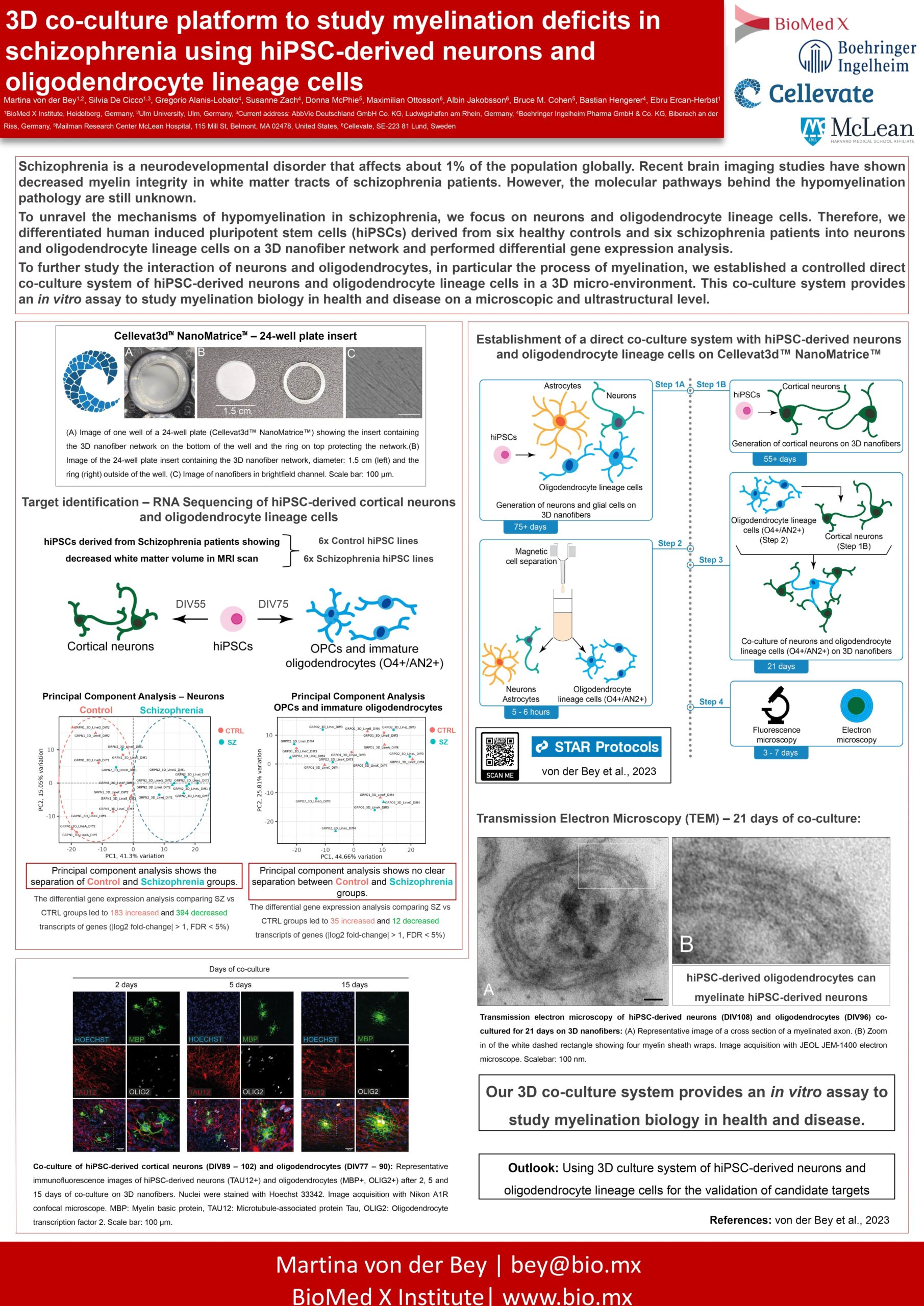
2. Poster: 3D co-culture platform to study myelination deficits in schizophrenia using hiPSC-derived neurons and oligodendrocyte lineage cells
**There is an error in the protocol . The thickness of the scaffold is 20um not 100um. The scaffold used is the same as catalog# 100-15 24-well plate Removable scaffold**
Ready to Use Models

Neuro
We have developed a scaffold for the culture of iPSCs to study psychiatric diseases. Neurons and astrocytes mature faster in a 3D culture compared to conventional 2D cultures, and the co-cultures can be maintained for several weeks without affecting cell viability. Initial staining indicates that neurons develop a very dense neuronal network, and the surrounding astrocytes are in close interaction with them. Furthermore, in collaboration with BioMed X, we developed a 3D co-culture protocol for studying myelination.
Custom Models

1. Breast Cancer
In collaboration with Lund University, we have developed a scaffold for the co-culture of cells used in breast cancer research. We have successfully demonstrated that various cell types, both malignant and normal, thrive in the 3D fiber unit. We are currently exploring this model for the co-culturing of cancer cells and different types of stromal cells. We anticipate that this model can be utilized to create a 3D tumor ex vivo platform suitable for screening therapeutic compounds, which will reduce the number of animals used in cancer research.

2. Pancreatic Cancer
In collaboration with Swedish CRO, we have developed a scaffold for stable, long-term co-culture of pancreatic cancer and stromal cells. Their model for pancreatic cancer co-cultures, a traditional submerged 2D system, had performance issues. Limited space resulted in problematic contact inhibition, and the system became over-confluent around 4 DIV, which did not allow sufficient time to develop a disease-relevant culture. The model is currently being used to further investigate drug sensitivity and cytotoxicity.
Customization
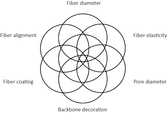
Cellevate uses networks of nanofibers to mimic different types of tissue. Our strength lies in our unique technology, combined with years of know-how and expertise, that allows us to fine tune the parameters of the nanomaterials we create and offer our customers tailor made solutions optimized for your specific applications.
Specification
- Material: Fully synthetic polycaprolactone (PCL)
- Manufacturer: Cellevate
Pricing
Cellevat3d™ NanoMatrice™ Neuro
- Scaffold for culture of iPSCs for studying psychiatric disease
12 well plate
Non-removable
- SKU: 100-09
- Price: $255.00
Removable
- SKU: 100-11
- Price: $255.00
24 well plate
Non-removable
- SKU: 100-13
- Price: $270.00
Removable
- SKU: 100-15
- Price: $270.00
48 well plate
Non-removable
- SKU: 100-17
- Price: $290.00
Removable
- SKU: 100-19
- Price: $290.00
96 well plate
Non-removable
- SKU: 100-19
- Price: $305.00
Cellevat3d™ NanoMatrice™
Breast Cancer
- Scaffold for co-culture of breast cancer research
This product is custom-made only.
Please contact us for a quote
Cellevat3d™ NanoMatrice™ Pancreatic Cancer Platform
- Scaffold for stable, long-term co-culture of pancreatic cancer and stromal cells
This product is custom-made only.
Please contact us for a quote
References & Literature
von der Bey, M., De Cicco, S., Zach, S., Hengerer, B. & Ercan-Herbst, E. Three-dimensional co-culture platform of human induced pluripotent stem cell-derived oligodendrocyte lineage cells and neurons for studying myelination. STAR Protocols 4, 102164 (2023) doi: 10.1016/j.xpro.2023.102164.
Malakpour-Permlid, A., Buzzi, I., Hegardt, C., Johansson, F. & Oredsson, S. Identification of extracellular matrix proteins secreted by human dermal fibroblasts cultured in 3D electrospun scaffolds. Scientific Reports 11, 6655 (2021) doi: 10.1038/s41598-021-85742-0.
Atena Malakpour Permlid, Plaurent Roci, Elina Fredlund, Felicia Fält, Emil Önell, Fredrik Johansson, Stina Oredsson, Unique animal friendly 3D culturing of human cancer and normal cells, Toxicology in Vitro, Volume 60, 2019, Pages 51-60, ISSN 0887-2333, https://doi.org/10.1016/j.tiv.2019.04.022.
Dahlin L.B., Stenberg L., Johansson U.E., Johansson F. (2019) Traumatic Peripheral Nerve Injuries: Experimental Models for Repair and Reconstruction. In: Risling M., Davidsson J. (eds) Animal Models of Neurotrauma. Neuromethods, vol 149. Humana, New York, NY. https://doi.org/10.1007/978-1-4939-9711-4_9
Frost, H.K., Andersson, T., Johansson, S. et al. Electrospun nerve guide conduits have the potential to bridge peripheral nerve injuries in vivo. Sci Rep 8, 16716 (2018). https://doi.org/10.1038/s41598-018-34699-8
Zalis MC, Nylander M, Enjin A, Stensmyr MC, Johansson P, O’Carroll D and Englund Johansson U (2019). Assessing electrical activity of human neuronal cell culture networks grown on 3D fibrous scaffolds using different techniques: A comparative study. Conference Abstract: MEA Meeting 2018 | 11th International Meeting on Substrate Integrated Microelectrode Arrays.
https://www.frontiersin.org/10.3389%2fconf.fncel.2018.38.00048/event_abstract
Albin Jakobsson, Maximilian Ottosson, Marina Castro Zalis, David O’Carroll, Ulrica Englund Johansson, Fredrik Johansson, Three-dimensional functional human neuronal networks in uncompressed low-density electrospun fiber scaffolds, Nanomedicine: Nanotechnology, Biology and Medicine, Volume 13, Issue 4, 2017, Pages 1563-1573, ISSN 1549-9634, https://doi.org/10.1016/j.nano.2016.12.023.
Englund-Johansson, U. , Netanyah, E. and Johansson, F. (2017) Tailor-Made Electrospun Culture Scaffolds Control Human Neural Progenitor Cell Behavior—Studies on Cellular Migration and Phenotypic Differentiation. Journal of Biomaterials and Nanobiotechnology, 8, 1-21. http://dx.doi.org/10.4236/jbnb.2017.81001
Ottosson M, Jakobsson A, Johansson F (2017) Accelerated Wound Closure – Differently Organized Nanofibers Affect Cell Migration and Hence the Closure of Artificial Wounds in a Cell Based In Vitro Model. PLoS ONE 12(1): e0169419. https://doi.org/10.1371/journal.pone.0169419
Marina Castro Zalis, Sebastian Johansson, Fredrik Johansson, Ulrica Englund Johansson, Exploration of physical and chemical cues on retinal cell fate, Molecular and Cellular Neuroscience, Volume 75, 2016, Pages 122-132, ISSN 1044-7431, https://doi.org/10.1016/j.mcn.2016.07.006.
FOR RESEARCH USE ONLY, NOT FOR USE IN DIAGNOSTIC PROCEDURES
