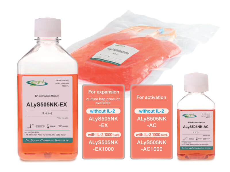
ALyS™505NK Series
Serum-free cell culture medium for activation and expansion of Human NK cells
Product Basics
ALyS505NK-AC and ALyS505NK-EX are serum-free, xeno-free cell culture media optimized for human natural killer (NK) cells derived from peripheral blood mononuclear cells (PBMCs). NK cells are crucial for innate immunity. Unlike T cells, they can damage virus-infected cells and cancer cells without prior sensitization.
Many applications necessitate the expansion of NK cells. However, in vitro cultivation of NK cells often results in a low expansion rate, an exhausted phenotype due to long-term expansion, and overgrowth of T cells if present in the initial culture. By employing ALyS505NK-AC for activation and ALyS505NK-EX for expansion, one can attain a highly pure, NK-rich population of cells in 14 days without the need for sorting with beads or flow cytometry. Both ALyS505NK-AC and ALyS505NK-EX are available with or without IL-2 (1000IU/mL).
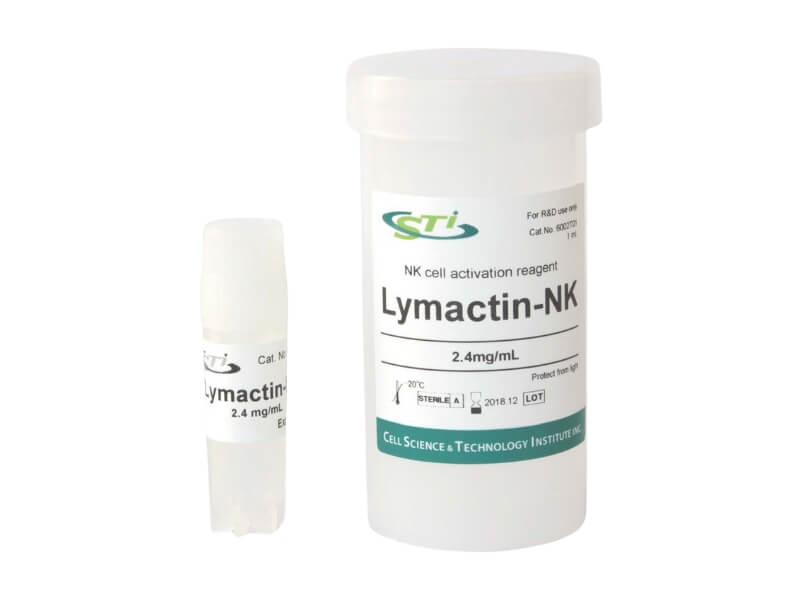
Artificial Serum is recommended for the primary culture derived from PBMC. Lymactin-NK, an anti-HER2 monoclonal antibody, is also recommended. It can activate human and animal peripheral blood lymphocytes by immobilizing them on culture vessels, thus inducing NK proliferation.
Key Features
- Serum-free (xeno-free) culture medium
- Facilitates efficient and high proliferation of NK cells.
- Enables the attainment of a higher ratio of NK cells to T cells
- Expanded NK cells exhibit cytotoxic activity at a low E/T ratio
- Available with or without IL-2 (1000IU/mL)
- Culture bag format for larger sizes is available for ALyS505NK-EX
- Optimal for NK cell therapy
Technical Information
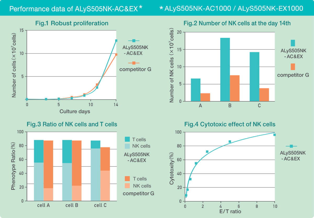
Robust Proliferation
As shown in Fig. 1, the proliferation of PBMC cultured in ALyS505NK-AC & EX over 14 days was higher than that in competitor G’s medium.
Highly efficient proliferation ratio of NK cells
In Fig. 2 and 3, PBMCs were collected from three donors and cultured with ALyS505NK-AC & EX and competitor G’s medium. As depicted in Fig. 2, the number of NK cells obtained on day 14 was significantly higher when cultured with ALyS505NK-AC & EX. Furthermore, as shown in Fig. 3, the ratio of NK cells to T cells was between 55-75% and 45-25% with ALyS505NK-AC & EX. In contrast, only 25-45% of NK cells were obtained after culturing in competitor G’s medium. Despite some noticeable variation among donors, ALyS505NK-AC & EX demonstrated highly efficient proliferation of NK cells and a high NK cell ratio.
Phenotype of NK cells are maintained
In Fig. 4, the cytotoxicity level of NK cells was evaluated by incubating them with K562 for four hours at varying E/T ratios. The NK cells cultured from ALyS505NK-EX exhibited cytotoxic activity even at low E/T ratios.
Specification
Scroll to right
| SKU | Product | Description | Size | Contents | Storage |
|---|---|---|---|---|---|
| 01600P02 | ALyS505NK-AC | IL-2 free (Activation) | 200ml | Bottle | 4°C |
| 01610P02 | ALyS505NK-AC1000 | IL-2 concentration 1,000lU/mL (Activation) | 200ml | Bottle | 4°C |
| 01400P10/01400C10 | ALyS505NK-EX | IL-2 free (Expansion) | 1L | Bottle/Culture Bag | 4°C |
| 01410P10/01410C10 | ALyS505NK-EX1000 | IL-2 concentration 1,000lU/mL (Expansion) | 1L | Bottle/Culture Bag | 4°C |
- Manufactured by: Cell Science and Technology Institute
Pricing
ALyS™505NK Series
- Serum-free cell culture medium for activation and expansion of Human NK cells
Activation
IL-2 Free
- SKU: 01600P02
- Size: 200ml (Bottle)
- Price:
$111.00→ $90.00
/
IL-2 concentration 1,000lU/mL
- SKU: 01610P02
- Size: 200ml (Bottle)
- Price:
$150.00→ $121.00
Expansion
IL-2 Free
- SKU: 01400P10
- Size: 1L (Bottle)
- Price:
$167.00→ $135.00
- SKU: 01400C10
- Size: 1L (Culture Bag)
- Price: Contact us
/
IL-2 concentration 1,000lU/mL
- SKU: 01410P10
- Size: 1L (Bottle)
- Price:
$283.00→ $230.00
- SKU:01410C10
- Size: 1L (Culture Bag)
- Price: Contact us
Complement Products
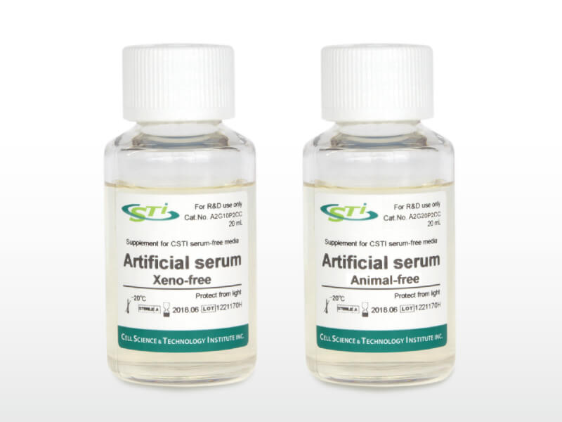
- Complement for Serum Free Medium
Animal Free
- SKU: A2G20P2C
- Size: 20ml
- Price: $946.00
References
- Zhou, Z., Li, T., Li, J., Lin, W. & Zheng, Q. Exosomal transfer of HCC-derived miR-17-5p downregulates NK cell function by targeting RUNX1-NKG2D axis. International Immunopharmacology 136, 112361 (2024) doi: 1016/j.intimp.2024.112361.
- Hong, S. D. et al. Trastuzumab-Mediated Antibody-Dependent Cell-Mediated Cytotoxicity (ADCC) Enhances Natural Killer Cell Cytotoxicity in HER2-Overexpressing Ovarian Cancer. International Journal of Molecular Sciences 25, (2024) doi: 3390/ijms252111733.
- Kim, S.-H. et al. Enhancement of the Anticancer Ability of Natural Killer Cells through Allogeneic Mitochondrial Transfer. Cancers 15, (2023) doi: 3390/cancers15123225.
- He, L. et al. Metabolic Reprogramming of NK Cells by Black Phosphorus Quantum Dots Potentiates Cancer Immunotherapy. Advanced Science 10, 2202519 (2023) doi: 1002/advs.202202519.
- Leem, G. et al. Safety and Efficacy of Allogeneic Natural Killer Cells in Combination with Pembrolizumab in Patients with Chemotherapy-Refractory Biliary Tract Cancer: A Multicenter Open-Label Phase 1/2a Trial. Cancers 14, (2022) doi: 3390/cancers14174229.
- Mikawa, S., Matsuda, A., Kamemori, Y., Asanuma, S. & Kitagawa, H. Enhancement of natural killer cell activity by oral administration of a fermented soybean product in dogs. Open Veterinary Journal 11, 394–400 (2021) doi: 5455/OVJ.2021.v11.i3.10.
- Jung, D. et al. Ex vivo expanded allogeneic natural killer cells have potent cytolytic activity against cancer cells through different receptor-ligand interactions. Journal of Experimental & Clinical Cancer Research 40, 333 (2021) doi: 1186/s13046-021-02089-0.
- Chang, J. et al. Diffracted X-ray blinking measurements of interleukin 15 receptors in the inner/outer membrane of living NK cells. Biochemical and Biophysical Research Communications 556, 53–58 (2021) doi: 1016/j.bbrc.2021.03.144.
- Liu, X., Sun, T., Ge, Q. & Zhu, J. Construction of Novel BispecificSingle-Domain Antibodies (BiSdAbs) with Potent Antiangiogenic Activities. Pharmaceutical Fronts 2, e64–e76 (2020) doi: 1055/s-0040-1708527.
- Nakamura, Y. et al. Natural killer cells impede the engraftment of cardiomyocytes derived from induced pluripotent stem cells in syngeneic mouse model. Scientific Reports 9, 10840 (2019) doi: 1038/s41598-019-47134-3.
- Lai, H. et al. Selenium-containing ruthenium complex synergizes with natural killer cells to enhance immunotherapy against prostate cancer via activating TRAIL/FasL signaling. Biomaterials 219, 119377 (2019) doi: 1016/j.biomaterials.2019.119377.
- Jung, I. H. et al. In Vivo Study of Natural Killer (NK) Cell Cytotoxicity Against Cholangiocarcinoma in a Nude Mouse Model. In Vivo 32, 771 (2018) doi: 21873/invivo.11307.
- Yu, H. et al. Large scale ex vivo expansion of clinical‑grade effector cells for adoptive immunotherapy. Exp Ther Med 14, 5678–5686 (2017) doi: 3892/etm.2017.5228.
- Niu, C. et al. Low-dose bortezomib increases the expression of NKG2D and DNAM-1 ligands and enhances induced NK and γδ T cell-mediated lysis in multiple myeloma. Oncotarget 8, 5954–5964 (2017) doi: 18632/oncotarget.13979.
- Singh, T. D., Lee, J. & Jeon, Y. H. Noninvasive Imaging of Natural Killer Cell-Mediated Apoptosis in a Mouse Tumor Model. in Natural Killer Cells: Methods and Protocols (ed. Somanchi, S. S.) 297–306 (Springer New York, 2016). doi: 1007/978-1-4939-3684-7_25.
- Shi, X. et al. Epigenetic suppression of the antitumor cytotoxicity of NK cells by histone deacetylase inhibitor valproic acid. Am J Cancer Res 6, 600–614 (2016)
Other Documents
Related Products
FOR RESEARCH USE ONLY, NOT FOR USE IN DIAGNOSTIC PROCEDURES