Exciting news! A groundbreaking study using BIOMIMESYS® for breast cancer metastasis research has been published in the Experimental Hematology & Oncology Journal. The study, titled “ProNGF promotes brain metastasis through TrkA/EphA2 induced Src activation in triple negative breast cancer cells” by Cicero, Trouvilliez, et al. (2023), emphasizes the role of Extracellular Matrix organ specificity in MDA-MB-231 breast cancer brain metastasis. Utilizing BIOMIMESYS® Brain and Liver matrices, which mimic the specific microenvironments of these organs, the study successfully replicated brain and liver metastasis behaviors in vitro, similar to those in mice. This not only proves BIOMIMESYS® as an effective tool for studying metastasis propagation but also offers a promising alternative to animal testing.
Source: https://ehoonline.biomedcentral.com/articles/10.1186/s40164-023-00463-6
| Product | Study model | Stiffness | Composition |
| Biomimesys Brain | Metastasis invasion | 0.1 kPa | Collagen IV |
| Biomimesys Liver | Metastasis invasion | 0.6 kPa | Collagen I and IV |
| Biomimesys Oncology | Developed cancer | 1.1 kPa | Collagen I |
Abstract
Background: Triple-Negative Breast Cancer is particularly aggressive, and its metastasis to the brain has a significant psychological impact on patients’ quality of life, in addition to reducing survival. The development of brain metastases is particularly harmful in triple-negative breast cancer (TNBC). To date, the mechanisms that induce brain metastasis in TNBC are poorly understood.
Methods: Using a human blood-brain barrier (BBB) in vitro model, an in vitro 3D organotypic extracellular matrix, an ex vivo mouse brain slices co-culture and in an in vivo xenograft experiment, key step of brain metastasis were recapitulated to study TNBC behaviors.
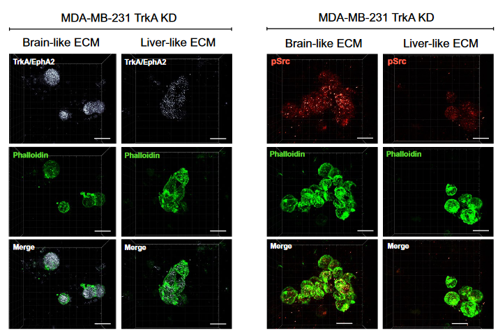
Results: In this study, we demonstrated for the first time the involvement of the precursor of Nerve Growth Factor (proNGF) in the development of brain metastasis. More importantly, our results showed that proNGF acts through TrkA independent of its phosphorylation to induce brain metastasis in TNBC. In addition, we found that proNGF induces BBB transmigration through the TrkA/EphA2 signaling complex. More importantly, our results showed that combinatorial inhibition of TrkA and EphA2 decreased TBNC brain metastasis in a preclinical model.
Conclusions: These disruptive findings provide new insights into the mechanisms underlying brain metastasis with proNGF as a driver of brain metastasis of TNBC and identify TrkA/EphA2 complex as a potential therapeutic target.